јҡ°ыҪMҝ—Пы»ҜіЈУГөДҺЧ·NГёөДЯx“с
 |
|
ЦұҪУҸДЙъОпуw«@ИЎөДҪMҝ—Ј¬Т»°гРиТӘҢўЖдПы»ҜіЙҶОӮҖјҡ°ыІЕДЬЯMРРуwНвЕарBЎЈЯ@·NЦұҪУҸДлxуwҪMҝ—«@өГөДјҡ°ыЈ¬ёьҪУҪьУЪЙъОпуwғИөДЙъ»о о‘BЈ¬ЗТЙъОпРФ оЙРОҙ°lЙъәЬҙуёДЧғЈ¬ТтҙЛФЪЛҺОпәYЯxЎўјҡ°ыТЖЦІЎўоҗЖч№ЩЕарBЎўД[БцСРҫҝөИұҠ¶аоIУтӮдКЬҡgУӯЎЈө«ҪMҝ—Пы»ҜЯ^іМЦРіЈУцөҪ¶а·NҶ–о}Ј¬АэИзПы»ҜІ»НкИ«Ўўјҡ°ыЛАНцВКёЯөИЎЈИзәОҝЛ·юЯ@Р©Ҷ–о}Ј¬ЖдҢҚПы»ҜГёөДЯx“сКЗкPжIЎЈ |
| ҒнБЛҪвҪMҝ—Пы»ҜЯ^іМЦРіЈУГөДГё°ЙЎ« |
|
1.ТИө°°ЧГё |
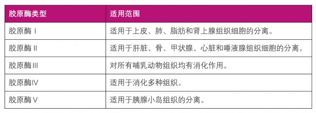 |
| ДzФӯГёоҗРНј°ЯmУГ·¶Үъұн |
|
3.НёГчЩ|ЛбГё |
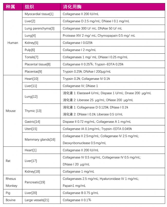 |
| ұнТ»ЈәХэіЈҪMҝ—Пы»ҜУГГёБРұн |
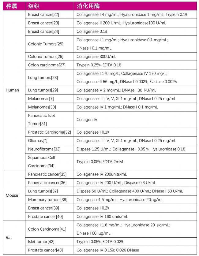 |
| ұн¶юЈәД[БцҪMҝ—Пы»ҜУГГёБРұн |
|
…ўҝјОД«I 1. Kasai-Brunswick TH., et al., (2017) Cardiosphere-derived Cells Do Not Improve Cardiac Function in Rats With Cardiac Failure. Stem Cell Res Ther. Feb 15;8(1):36. 2. Huch.M., et al., (2015) Long-term Culture of Genome-Stable Bipotent Stem Cells From Adult Human Liver. Cell. Jan 15;160(1-2):299-312. 3. Holt PG., et al., (1986) Extraction of Immune and Inflammatory Cells From Human Lung Parenchyma: Evaluation of an Enzymatic Digestion Procedure. Clin Exp Immunol. Oct;66(1):188-200. 4. Campbell AM., et al., (1993) Modulation of Eicosanoid and Histamine Release From Human Dispersed Lung Cells by Terfenadine. Allergy. Feb;48(2):125-9. 5. Sanchez-Romero N., et al., (2020) A Simple Method for the Isolation and Detailed Characterization of Primary Human Proximal Tubule Cells for Renal Replacement Therapy. Int J Artif Organs. Jan;43(1):45-57. 6. Ataollahi F., et al., (2014) New Method for the Isolation of Endothelial Cells From Large Vessels. Cytotherapy. Aug;16(8):1145-52. 7. Leelatian N., et al., (2017) Single cell analysis of human tissues and solid tumors with mass cytometry. Cytometry B Clin Cytom. Jan;92(1):68-78. 8. Roberts EG., et al., (2019) Evaluation of Placental Mesenchymal Stem Cell Sheets for Myocardial Repair and Regeneration. Tissue Eng Part A Jun;25(11-12):867-877. 9. Jankovic-Karasoulos T., et al., (2018) Isolation of villous cytotrophoblasts from second trimester human placentas. Placenta. Dec 15;74:55-58. 10. Tang YL., et al., (2007) A Novel Two-Step Procedure to Expand Cardiac Sca-1+ Cells Clonally. Biochem Biophys Res Commun. Aug 10;359(4):877-83. 11. Meyer J. ., et al., (2016) An optimized method for mouse liver sinusoidal endothelial cell isolation. Exp Cell Res. Dec 10;349(2):291-301. 12. Nakano H., et al., 1799 (2018) Isolation and Purification of Epithelial and Endothelial Cells from Mouse Lung. Methods Mol Biol.:59-69. 13. Jain R. ., et al., (2014) Isolation of thymic epithelial cells and analysis by flow cytometry. Curr Protoc Immunol. Nov 3;107:3.26.1-3.26.15. 14. Tran LS., et al., (2018) Isolation of Mouse Primary Gastric Epithelial Cells to Investigate the Mechanisms of Helicobacter Pylori Associated Disease. Methods Mol Biol. 1725:119-126. 15. De Clercq K., et al., (2017) Isolation of Mouse Endometrial Epithelial and Stromal Cells for In Vitro Decidualization. J Vis Exp Mar 2;(121):55168. 16. Sharon, Y., et al., (2013) Isolation of Normal and Cancer-Associated Fibroblasts from Fresh Tissues by Fluorescence Activated Cell Sorting (FACS). J Vis Exp 71, e4425. 17. Zhang Q., et al., (2016) Isolation and Culture of Single Cell Types from Rat Liver. Cells Tissues Organs. 201(4):253-67. 18. Al-Eisa A., (2017) IgA Enhances IGF-1 Mitogenic Activity Via Receptor Modulation in Glomerular Mesangial Cells: Implications for IgA-Induced Nephropathy. Kidney Blood Press Res.;42(3):391-397. 19. Githens S., et al., (1994) Isolation and Culture of Rhesus Monkey Pancreatic Ductules and Ductule-Like Epithelium Pancreas. Jan;9(1):20-31. 20. Caperna TJ., et al., (2011) Culture of porcine hepatocytes or bile duct epithelial cells by inductive serum-free media In Vitro Cell Dev Biol Anim. Mar;47(3):218-33. 21. Ataollahi F., et al., (2014) New method for the isolation of endothelial cells from large vessels. Cytotherapy. Aug;16(8):1145-52. 22. Widowati W., et al., (2018) Isolation, Characterization and Proliferation of Cancer Cells from Breast Cancer Patients. Acta Inform Med. Dec;26(4):240-244. 23. Beaupain R., et al., (1993) “Normal” breast cells adjacent to a tumor grown in long-term three-dimensional culture. In Vitro Cellular & Developmental Biology – Plant, 29(2): 100-104. 24. Leung C K., et al., (1982) Morphological and proliferative characteristics of human breast tumor cells cultured on plastic and in collagen matrix. In Vitro Cellular & Developmental Biology – Plant, 18(5): 476-482. 25. Chou J., et al., (2013) Phenotypic and Transcriptional Fidelity of Patient-Derived Colon Cancer Xenografts in Immune-Deficient Mice. PLOS ONE, 8(11). 26. Friedman E., et al., (1981) Tissue culture of human epithelial cells from benign colonic tumors. In Vitro Cellular & Developmental Biology – Plant, 17(7): 632-644. 27. Brattain M G., et al., (1983) Characterization of human colon carcinoma cell lines isolated from a single primary tumour. British Journal of Cancer, 47(3): 373-381. 28. Quatromoni J G., et al., (2015) An optimized disaggregation method for human lung tumors that preserves the phenotype and function of the immune cells. Journal of Leukocyte Biology, 97(1): 201-209. 29. Zhuang X., et al., (2015) Identification of novel vascular targets in lung cancer Br J Cancer. Feb 3; 112(3): 485–494. 30. Welte Y., et al., (2013) Patient Derived Cell Culture and Isolation of CD133+ Putative Cancer Stem Cells from Melanoma. Journal of Visualized Experiments. 31. Tillotson LG., et al., (2001) Isolation, Maintenance, and Characterization of Human Pancreatic Islet Tumor Cells Expressing Vasoactive Intestinal Peptide Pancreas. Jan;22(1):91-8. 32. Nakashiro K., et al., (2004) Phenotypic Switch from Paracrine to Autocrine Role of Hepatocyte Growth Factor in an Androgen-Independent Human Prostatic Carcinoma Cell Line, CWR22R. American Journal of Pathology, 165(2): 533-540. 33. Sheela S., et al., (1990) Angiogenic and invasive properties of neurofibroma Schwann cells. Journal of Cell Biology, 111(2): 645-653. 34. Sacks P G., et al., (1988) Establishment and characterization of two new squamous cell carcinoma cell lines derived from tumors of the head and neck. Cancer Research, 48(10): 2858-2866. 35. Kim M P., et al., (2009) Orthotopic and heterotopic generation of murine pancreatic cancer xenografts [J]. Nature Protocols, 4(11): 1670-1680. 36. Rasheed Z A., et al., (2010) Isolation of Stem Cells from Human Pancreatic Cancer Xenografts. Journal of Visualized Experiments. 37. Vaughan A E., et al., (2011) Lung Cancer in Mice Induced by the Jaagsiekte Sheep Retrovirus Envelope Protein Is Not Maintained by Rare Cancer Stem Cells, but Tumorigenicity Does Correlate with Wnt Pathway Activation. Molecular Cancer Research, 10(1): 86-95. 38. Liu X., et al., (2013) Nonlinear Growth Kinetics of Breast Cancer Stem Cells: Implications for Cancer Stem Cell Targeted Therapy. Scientific Reports , 3(1): 2473-2473. 39. Kazerounian S., et al., (2013) RhoB differentially controls Akt function in tumor cells and stromal endothelial cells during breast tumorigenesis. Cancer Research, 73(1): 50-61. 40. Mazzoleni S., et al., (2013) Gene Signatures Distinguish Stage-Specific Prostate Cancer Stem Cells Isolated From Transgenic Adenocarcinoma of the Mouse Prostate Lesions and Predict the Malignancy of Human Tumors. Stem Cells Translational Medicine, 2(9): 678-689. 41. Duarte S., et al., (2013) Preventive Cancer Stem Cell©\Based Vaccination Reduces Liver Metastasis Development in a Rat Colon Carcinoma Syngeneic Model. Stem Cells, 31(3): 423-432. 42. Gazdar A F., et al., (1980) Continuous, clonal, insulin- and somatostatin-secreting cell lines established from a transplantable rat islet cell tumor. Proceedings of the National Academy of Sciences of the United States of America, 77(6): 3519-3523. 43. Sharma N K., et al., (1999) A Novel Immunological Model for the Study of Prostate Cancer. Cancer Research, 59(10): 2271-2276. |
 |
В“ПөлҠФ’Јә40000 53055,0512 6832 5983
E-mailЈәpeprotech.infochina@thermofisher.com

- PCL-PVAc-PEGФЪ»оРФОпЯfЛНәНЙъОпІДБПЙПөД‘ӘУГғһ„Э
- PROTACЯBҪУЧУЦРЧчУГҷCЦЖј°өӘлsӯhөДҪYҳӢғһ„ЭЕc№ҰДЬМШРФ
- OK432ФЪД[БцәНГвТЯСРҫҝЦРөДЧчУГҷCЦЖј°‘ӘУГғһ„Э
- иFХ{ЛШHepcidin-25ЧчһйиFҙъЦxәНГвТЯ·ҙ‘ӘкPжIХ{№қТтЧУөД№ҰДЬәНЧчУГ
- МЗө°°ЧFibronectinФЪјҡ°ыЕарBЦРөД№ҰДЬј°ЧчУГ
- TDP-43өДҪYҳӢЕc№ҰДЬј°ФЪқuғц°Y(ALS)өИЙсҪӣНЛРРРФјІІЎЦРөДЧчУГҷCЦЖ
- DNP-BSA(2,4-¶юПх»щұҪЕјВ“ЕЈСӘЗе°Чө°°Ч)ФЪГвТЯҢWСРҫҝЕcҷzңyЦРөДЧчУГ
- AbMole LPSЈЁЦ¬¶аМЗЈ©ФЪГвТЯјӨ»оЎў„УОпФмДЈөИ·ҪГжөД‘ӘУГј°°ёАэ·ЦПн
- MCEЦРҮшҢў”yФҮ„©®aЖ·ББПаГАҮшЦҘјУёз2025 AACRДк•ю
- 2024Дк"MCEЦРҮшЙъГьҝЖҢWСРҫҝҙЩЯMӘ„"ФuӘ„ҪY№ы№«Іј
- МХРgЙъОпЦұІҘоAёжЈәКЬуwЛҺАнҢWЕcЛҺОп°l¬FСРҫҝ
- °ІҪЭӮҗГвТЯҪM»Ҝ·ЁPD-L1ҷzңyФҮ„©әР«@өГҡWГЛIVDRХJЧC
- өВҮшMassive PhotonicsНЖіц¶аҝоDNA-PAINTёчПөФҮ„©әР
- МмВЎЧФЦчСР°lHCVәЛЛбҷzңyФҮ„©әР«@ЕъЛҺұOҫЦИэоҗNMPA
- МmІ©АыөВНЖіцРВЖ·ёЯұЈХжҝмЛЩ”UФц PCR mixФҮ„©әР
- IPHASEСыДъ№Іё°BIOCHINA2025өЪК®ҢГТЧЩQЙъОп®aҳIҙу•ю