賽多利斯空氣浮游菌采集儀MD8采集流感病毒(H3N2)
德國賽多利斯公司空氣浮游菌采集儀MD8 采用膜過濾方法采集空氣中流感病毒(H3N2)
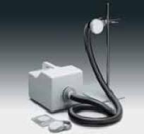
空氣浮游菌采集儀MD8由空氣浮游菌采樣器和拋棄型凝膠膜組成。該系統通常被用于定量檢測的空氣浮游菌,主要是在灌裝線上的Class A級無菌區域(級別依據“歐盟GMP指南”分類),隔離間或吹塑-灌裝-封口無菌包裝機器。
高達8 m3/h的空氣流速能夠等速采樣。
通常在層流罩和過濾1 m3 空氣的速度非常快(不到8分鐘)。
該采樣頭可與空氣采樣器分開放置,可進行遠程采樣。
凝膠膜和MD 8 空氣采樣器配合使用可提供以下特點和好處:
-"絕對"的截留率( Bac. sub. niger spores 9 9.9995%,T3 phages 99.94 %) 。
-在采樣過程中,凝膠膜能夠保持微生物的活性
-凝膠膜是完全水溶性的。因此,一個樣品中的微生物可以在幾種不同的培養基中培養,或可測出低和高的細菌數。樣品不受空氣抑制劑的影響。
-凝膠膜的溶解性是采集病毒的一個必要條件。
Detecting Airborne Influenza Virus A (H3N2) in a Children's Polyclinic in Greifswald, Germany, by Collecting Virus Using the Water-soluble 12602 Sartorius Gelatin Membrane Filter
Introduction
Although the impinger and impaction collectors based on the impaction principle have long
ago proved their reliability for the collection of microbial aerosols, filters only recently
began to receive increased attention with the development of the water-soluble gelation filter technique and the wide spectrum of applications offered by such filters only for collection of bacterial aerosols; however – after Goettingen-based Sartorius AG had started commercial-scale manufacture of such gelatin filters.
Until now, the value of filters for detection and collection of virus aerosols has been considered limited, and the filters were thought to be useful for collecting stable, resistant viruses, if at all. There has been very little experimental data about this until recently.
Studies done by the Jaschhof team with experimentally pro-duced static aerosols of Tl and T3 coli phages and influenza viruses, strain A/PR/8/34 (H1N1) using the Sartorius Gelatin Membrane Filter (12602) yielded a constant collection efficiency for these viruses, even under conditions involving high inlet velocities (up to 1.6 m/s max.) and an extended collection period (15 minutes max.).
The high resistance of the gelatin filter to mechanical stress and the proven stability of the influenza virus were the reasons for testing the effectiveness of this method in practice during the seasonal rise in morbidity for acute respiratory diseases (ARD) at the beginning of 1990 (3rd – 5th week). This study builds upon results obtained from tests with experimentally produced virus and phage aerosols.
Air samples were taken over a period of 3 days in the waiting room of a Greifswald children’s polyclinic and the water-soluble filter material was then used in virus cultivating experiments. Experiments for cultivating the virus isolated from nasal secretions obtained by aspiration from the children in the waiting room, who had a fresh case of ARD, were performed in parallel.
Materials and Methods
Location Studied:
Waiting room of a Greifswald children's clinic (approx 120 m3) frequented by a maximum of 57 children accompanied by a parent over a period of 2 hours (Jan. 24, 1990). Temperature: 25°C, 60% relative humidity.
Experimental/Test Materials:
Nasal secretions from children with a fresh case of ARD. Gelatin membrane filter, 12602, after 1.8 m3of room air was filtered through it. It was then sealed in a polyethylene bag along with 5 ml of an isotonic buffer for cell cultivation at pH 7.4 with 0.5% yeast extract (Difco), penicillin G (400 lUs/ml) and streptomycin (400 µg/ml). All samples were processed within 4 hrs (with storage in between in a cooling chamber).
Air sampling:
3 days of air filtration with each sample consisting of 1.8 m3 of air per filter (120 I/min) during a collection period of 15 min through a Sartorius Gelatin Filter (126 02-050).
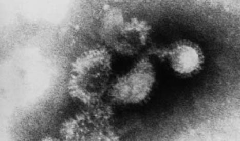
Scanning electron micograph of the influenza virus A/PR/8/34 (H0N1).
Viruses from a culture of an 11-day old embryonic chicken egg and purified with a saccharose density gradient. Micrograph: Herrmann/Fischer
Viruses from a culture of an 11-day old embryonic chicken egg and purified with a saccharose density gradient. Micrograph: Herrmann/Fischer
Virological Analyses:
Nasal secretions: Hank's solution (3 ml per sample) was added and the samples were centrifuged for 30 min (3,000 rpm), each given an additive of 10 µl antibiotic solution/ml of supernatant, and incubated for 1 hr at room temperature. The amnion of 10-day old incubated egges was inoculated with these specimens and incubated for 3 days at 33°C; 3 blind subcultures were prepared; HAT was used to detect the viruses.
Air samples: The filter was liquified for 5 min at 37°C in a water bath; an antibiotic solution
as described above was added to the solution which was used to inoculate incubated eggs; FL cell and ascites tumor cells were then cultured; the viruses were detected according to the usual techniques by HAT, CPE, and HAdT.
The influenza virus isolated from the nasal secretions and air samples were typed and identified by cross titration in HAHT with rabbit immune sera against the influenza virus A/Greifswald/1/89 (H1N1) and A/Greifswald/1/90(H3N2) and antigens from the influenza virus A/Sichuan/2/87 (H3N2) and influenza virus A/Greifswald/2/88 (H3N2).
The v-RNA and virus protein patterns were determined by PAGE.
Results and Discussion:
From the experimental material taken on Jan. 24, 1990, three types of viruses isolated from the nasal secretions and an influenza virus A isolated from an air sample were successfully inoculated in the incubated embryonated eggs during the third transfer for subculturinq (influenza virus A/Greifswald/2/90 [H3N2]). HAT showed that the antigens of the isolated viruses and those of the variants A/Greifswald/ 2/88 and A/Sichuan/2/87 were largely related and did not exhibit any differences from a strain which was cultivated from a nasal secretion specimen taken from a patient who was in the room at the time of air sampling using filters (influenza virus A/ Greifswald/1/90 [H3N2]).
The migration rates of the isolated viruses' proteins (HA, NP, NS and M) and all bands of their v-RNA following PAGE (poly-acrylamide gel electrophoresis) matched to a high degree (Fig. 1).
Attempts to isolate further viruses from air samples using cell cultures were not successful.
The fact that the air samples were taken in the pre-epidemic phase of the ARD morbidity
dynamics demonstrates the effectiveness of the filter collection method used in detecting
airborne influenza viruses.
Seventy-five percent of the children (> 8 years old) present on the day of the studies had a respiratory infection, but only 40% of these children at most showed acute catarrhal symptoms (coughing, sneezing, runny nose). The detection of the influenza virus A in the air of a children’s clinic waiting room, during a period of only a slight increase in seasonal morbidity from acute respiratory diseases in the region, remarkably confirms* the results obtained from using experimen-tally produced static aerosols of coli phages and influenza virus A.
This verifies, for the first time the suitability of the gelatin filter in practice for the collection of respiratory viruses from the air. * (see Sartorius ApplicationNotes Publication No. SLF4028-e95092)
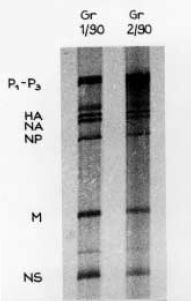
Figure 1:
Separation of the v-RNA in polyacrylamide gel electrophoresis (PAGE) from the influenza virus A/Greifswald/1/90 (H3N2) (isolated from patient specimens) and the influenza virus A/Greifswald/2/90 (collected and isolated from the air.) 3.5% polyacrylamide gel in tris-acetate EDTA buffer.
Separation of the v-RNA in polyacrylamide gel electrophoresis (PAGE) from the influenza virus A/Greifswald/1/90 (H3N2) (isolated from patient specimens) and the influenza virus A/Greifswald/2/90 (collected and isolated from the air.) 3.5% polyacrylamide gel in tris-acetate EDTA buffer.
Outlook
Considering the uncomplicated method of air filtration involving comparatively less work, this principle using the gelatin membrane filter is bound to receive increased recognition in helping to answer a multitude of questions about the production, stability and spread of infectious virus aerosols (e.g., involved in genetic engineering techniques).
Supplementary issue of the condensed text version of a poster displayed during the Annual Conference of the Society of Virology, Potsdam March 19-23, 1991).
Names and addresses of the authors:
Dr. Helmut Jaschhof Ph. D. in Natural Sciences (Dr. rer. nat.) Gerhard-Katsch-Str. 1417489 Greifswald, Germany
Dr. E.-J. Finke, M.D. Bundeswehrkrankenhaus Berlin Scharnhorststr. 1310115 Berlin 1040, Germany
Dr. B. Herrmann Ph. D. in Natural Sciences (Dr. rer. nat.)
und A. Reinecke Institut für Medizinische Mikrobiologie und Epidemiologie (lnstitute of Medical Microbiology and Epidemiology) E.-M.-Arndt-Universität Greifswald, Martin-Luther-Straße 6,17489 Greifswald, Germany
德國賽多利斯公司
http://www.sartorius-stedim.com.cn
地址:北京市順義區空港工業區B區裕安路33號
郵編:101300
電話:86-10-80426516
傳真:86-10-80426580
郵箱:XiaoDan.Yan@Sartorius-Stedim.com
- 新型FluorCam葉綠素熒光儀上市,助力設備以舊換新
- 中喬新舟推出HUVEC轉染試劑,獨家破解基因轉染壁壘
- 徠卡即將推出Visoria B/M/P三款新型正置手動顯微鏡
- 博鷺騰2025年合作伙伴大會圓滿成功并發布多款新品
- 上海優耳推出動物行為獎勵飼料,助力實驗動物研究
- 韓國LCI推出新一代組織透明化澄清液 Opti Mount
- 奧豪斯推出全新DEFENDER系列 DF2500電子平臺秤
- 銳視科技重磅發布全鏈條動物影像和生物輻照產品矩陣
- EVIDENT推出EviStar奧之星ES818顯微互動教學系統
- 艾普拜qMate mini新品上市,全面布局分子檢測產品線
- 勤翔新品IVScope8000M全光譜小動物活體成像系統試用
- 打破邊界精準進化,NEOSCAN推出NXL臺式大倉顯微CT
- 德國IKA歐洲之星EUROSTAR系列頂置攪拌器煥新升級
- 賽默飛蘇州TSX通用系列國產化超低溫冰箱震撼上市
- 勤翔發布新品IVScope 8000Pro小動物活體成像系統
- 洛科儀器誠摯邀您參加2025環境分析研討會
- 沃德精準亮相第26屆中國環博會,共赴環保新征程
- 萊闊邀您參加Harvesting Carbon 作者見面會
- ECHOES試驗揭秘冷流雪,萊比信設備助力氣象新突破
- 2025第五屆新疆國際畜牧業博覽會通知
- UVP系統等助力索邦大學全球浮游動物生物量精準評估
- 2025上海環境監測展暨上海儀器儀表展通知
- 2025中國(成都)畜牧業博覽會即將盛大開幕
- 研討會邀請:下一代渦度相關碳水通量測量解決方案
- 水德成功參展第七屆海洋環境開放科學大會XMAS 2025
- WD-SZ1000植物生長監測系統在山東某森林公園安裝
- 第十三屆生物質能源與有機固廢資源化利用論壇通知
- 水德攜ZooSCAN系統亮相第十屆全國海洋生態研討會
- LI-COR研討會:蒸散發ET數據自動化解決方案(12.5)
- 上海瑾瑜應邀參加2024中國海洋經濟博覽會
Copyright(C) 1998-2025 生物器材網 電話:021-64166852;13621656896 E-mail:info@bio-equip.com


